.gif)
Anatomy of the Lingual Nerve
The lingual nerve is a branch of the mandibular division of the trigeminal nerve supplying the anterior two thirds of the tongue and responding to stimuli of pressure, touch, and temperature (Image #1 & 2).
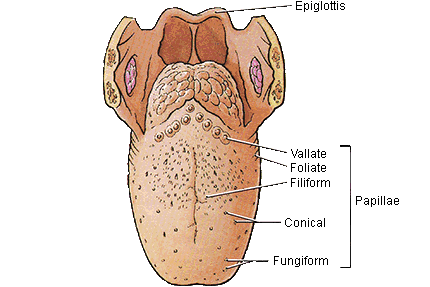
Image #1 : Tongue Anatomy
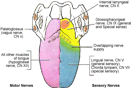
Image #2: Lingual Nerve Innervation
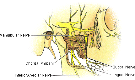
There is a lingual nerve for the right side of the tongue and one for the left side. The lingual nerve also carries a branch of the facial nerve called the chorda tympani which splits off the lingual nerve before the tongue is innervated and provides the sensation of taste to the anterior (front) two-thirds of the tongue. The mandibular nerve (V3) is the largest of the three branches of the trigeminal nerve. We are interested in the posterior division of the mandibular nerve which separates into the lingual nerve and the inferior alveolar nerve (Image #3). The lingual nerve runs to the medial (inside of the mandible or lower jaw) and next to the inferior alveolar nerve (Image #2) as they move towards the ramus of the mandible.
Image #3: Mandibular Nerve with the lingual nerve and the inferior alveolar nerve
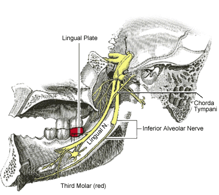
The lingual nerve takes the inside track next to the mandible and runs next to the third molar (wisdom tooth or teeth No. 17 on the left and No. 32 on the right -Image #3) on the lingual side (tongue side) of the third molar(Image #5).
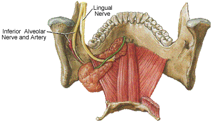
Image #4: Path of the lingual nerve which tracks to the inside of the mandible (lower jaw)
There is a boney barrier between the third molar and the lingual nerve called the lingual plate (Image #5) which the lingual nerve runs along on the lingual side (tongue side).
Image #5: Lingual nerve in relation to the lingual plate and third molar.
After passing the third molar, the lingual nerve runs below tongue, sending nerve branches up to the anterior (front) two thirds of the tongue (See Image #2 above which demonstrates the area of the tongue which the lingual nerve innervates in blue).The lingual nerve also carries nerve fibers that are not part of the trigeminal nerve, including the chorda tympani nerve (a branch of the facial nerve or cranial nerve VII), which provides special sensation (taste) to the anterior 2/3 part of the tongue. It is also important to keep in mind the inferior alveolar bundle in relationship to the third molar which can also be injured during the third molar extraction (See Inferior alveolar nerve damage following removal of mandibular third molar teeth. A true close relationship between the third molars and the mandibular canal increases the risk of injury to the inferior alveolar nerve, and an accurate evaluation of the relationship is essential to avoid the risk of surgery. Dentists should be aware of the limitations of the radiographic m
arkers of panoramic radiography and should consider the more detailed imaging of CBCT in specific cases in which the radiographic findings raise concerns of potential injury to the inferior alveolar nerve due to the close relationship between the third molar and the mandibular canal (See "Correlation of the radiological predictive factors of inferior alveolar nerve injury with cone beam computed tomography findings" and "Diagnostic Accuracy of PanoramicRadiography in Determining Relationship Between Inferior Alveolar Nerve and Mandibular Third Molar".
